Calculus prostatitis is accompanied by increased urination, dull pain in the lower abdomen and perineum, erectile dysfunction, the presence of blood in the seminal fluid, and prostatorrhea. Calculus prostatitis can be diagnosed using digital examination of the prostate, ultrasound of the prostate gland, urographic examination, and laboratory examinations. Conservative therapy for calculous prostatitis is carried out with the help of drugs, herbal remedies, and physiotherapy; If these measures are ineffective, stone destruction with a low-intensity laser or surgical removal is indicated.
General information
Calculus prostatitis is a form of chronic prostatitis, accompanied by the formation of stones (prostatoliths). Calculous prostatitis is the most common complication of the long-term inflammatory process in the prostate gland, which should be addressed by specialists in urology and andrology. During preventive ultrasound examinations, prostate stones are detected in 8. 4% of men of various ages. The first age peak in the incidence of calculous prostatitis occurs at the age of 30-39 years and is due to the increase in cases of chronic prostatitis caused by STDs (chlamydia, trichomoniasis, gonorrhea, ureaplasmosis, mycoplasmosis, etc. ). In men aged 40-59 years, calculous prostatitis, as a rule, develops against the background of prostate adenoma, and in patients over 60 years old it is associated with a decrease in sexual function.
The cause of prostatitis is calculus
Depending on the cause of formation, prostate stones can be true (primary) or false (secondary). Primary stones are initially formed directly in the acini and ducts of the gland, secondary stones migrate into the prostate from the upper urinary tract (kidney, bladder or urethra) if the patient has urolithiasis.
The development of calculous prostatitis is caused by congestive and inflammatory changes in the prostate gland. Impaired emptying of the prostate gland due to BPH, irregular or lack of sexual activity, and a sedentary lifestyle. Against this background, the addition of moist infections to the genitourinary tract leads to blockage of the prostate duct and changes in the nature of prostate secretion. On the other hand, prostate stones also support chronic inflammatory processes and secretion stagnation in the prostate.
In addition to stagnation and inflammatory phenomena, urethro-prostatic reflux plays an important role in the development of calculous prostatitis - pathological reflux of a small amount of urine from the urethra into the prostate duct during urination. At the same time, the salt contained in the urine crystallizes, thickens and, over time, turns into stones. Causes of urethro-prostatic reflux can be narrowing of the urethra, trauma to the urethra, atony of the prostate and seminal tubercles, previous transurethral resection of the prostate gland, etc.
The morphological core for prostatic calculus is amyloid bodies and desquamated epithelium, which gradually "grows" with phosphates and calcareous salts. Prostate stones are located in cystically distended acini (lobules) or in the excretory duct. Prostatoliths are yellowish, spherical in shape, and vary in size (on average from 2. 5 to 4 mm); may be single or multiple. In terms of its chemical composition, prostate stones are the same as bladder stones. With prostatitis, calculi, oxalate, phosphate and urinary stones are most often formed.
Symptoms of calculous prostatitis
The clinical manifestations of calculous prostatitis generally resemble the chronic inflammatory process of the prostate. The main symptom in the clinic of calculus prostatitis is pain. The pain is dull, painful in nature; localized in the perineum, scrotum, above the pubis, sacrum or coccyx. Exacerbating painful attacks may be associated with defecation, sexual intercourse, physical activity, prolonged sitting on hard surfaces, prolonged walking or bumpy driving. Calculous prostatitis is accompanied by frequent urination, sometimes with complete urinary retention; hematuria, prostatorrhea (leakage of prostate secretions), hemospermia. Characterized by decreased libido, weak erections, impaired ejaculation, and painful ejaculation.
Endogenous prostate stones can remain in the prostate gland for a long time without symptoms. However, a prolonged chronic inflammatory process and associated calculous prostatitis can lead to the formation of prostate abscess, the development of vesiculitis, atrophy and sclerosis of glandular tissue.
Diagnosis of calculous prostatitis
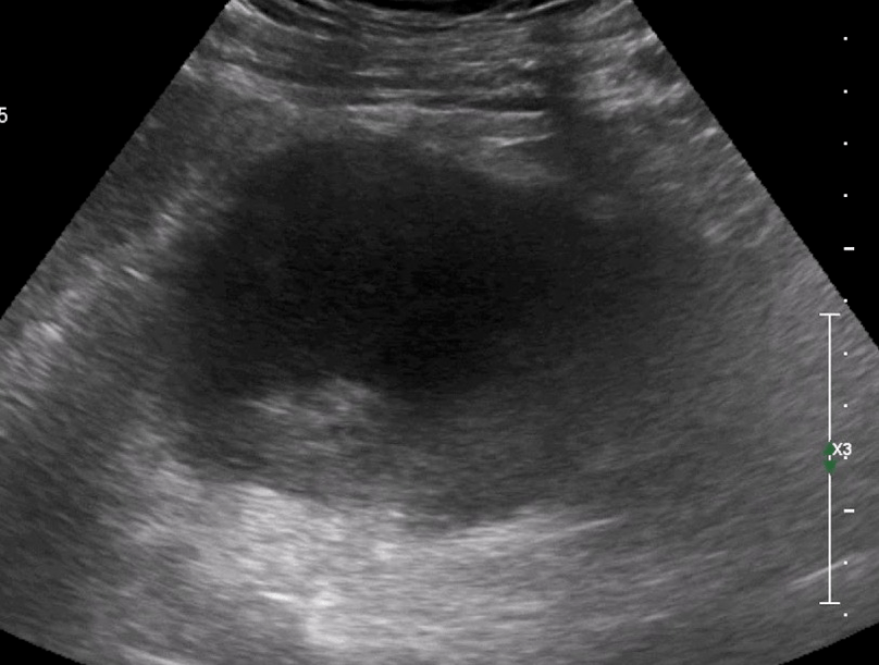
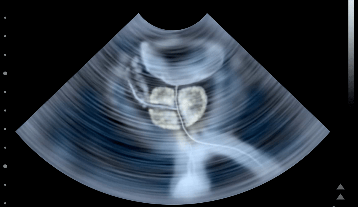
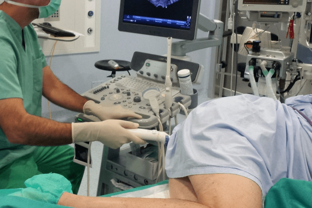
To establish the diagnosis of calculous prostatitis, consultation with a urologist (andrologist), evaluation of existing complaints, and physical and instrumental examination of the patient is required. When performing a digital examination of the prostate rectum, a lumpy stone surface and a kind of crepitus are determined by palpation. Using transrectal ultrasound of the prostate gland, stones are detected in the form of a hyperechoic formation with a clear acoustic track; its location, quantity, size and structure are explained. Sometimes urography, CT and MRI of the prostate are used to detect prostatolith. Exogenous stones are diagnosed with pyelography, cystography and urethrography.
Instrumental examination of patients with calculous prostatitis is complemented by laboratory diagnostics: examination of prostate secretions, bacteriological culture of urethral discharge and urine, PCR examination for scraping for sexually transmitted infections, biochemical analysis of blood and urine, determination of prostate-specific antigen levels, sperm biochemistry, ejaculate culture, etc.
During examination, calcified prostatitis is differentiated from prostate adenoma, tuberculosis and prostate cancer, bacterial and chronic abacterial prostatitis. In calculous prostatitis that is not associated with prostate adenoma, the volume of the prostate gland and the PSA level remain normal.
Treatment of calculous prostatitis

Uncomplicated stones in combination with chronic inflammation of the prostate gland require conservative anti-inflammatory therapy. Treatment of calculous prostatitis includes antibiotic therapy, non-steroidal anti-inflammatory drugs, herbal drugs, physiotherapeutic procedures (magnetic therapy, ultrasound therapy, electrophoresis). In recent years, low-intensity lasers have been successfully used to destroy prostate stones non-invasively. Prostate massage for patients with calculous prostatitis is strictly prohibited.
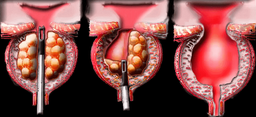
Surgical treatment of calculous prostatitis is usually required in cases of complicated disease, its combination with prostate adenoma. When a prostate abscess is formed, the abscess is opened, and along with the outflow of pus, the passage of stones is also observed. Sometimes mobile exogenous stones can be pushed instrumentally into the bladder and subjected to lithotripsy. The removal of fixed stones of large size is performed in the process of the perineal or suprapubic part. When calculous prostatitis is combined with BPH, the optimal method of surgical treatment is adenomectomy, prostate TUR, prostatectomy.
Treatment of calculous prostatitis
Calculous prostatitis is an inflammation of the prostate gland, complicated by the formation of stones. This type of prostatitis is the result of long-term chronic inflammation of the prostate. The disease is accompanied by frequent urination, painful pain in the lower abdomen and perineum, erectile dysfunction, and the presence of blood in the ejaculation.
The cause of this disease
Calculous is a type of chronic prostatitis characterized by stone formation. This disease is often a complication of a long-term inflammatory process in the prostate. Against the background of chronic inflammation under the influence of negative internal and external factors, the secretion stagnates, which over time crystallizes and turns into stones.
In addition to congestion and inflammatory phenomena, urethro-prostatic reflux, which is characterized by the pathological reflux of a small amount of urine from the urethra into the duct of the prostate gland during urination, plays a major role in the development of calculous prostatitis. The salt contained in the urine gradually crystallizes and over time turns into solid stones. Common causes of annual prostatic reflux:
- urethral injury;
- atony of the prostate gland and seminal tubercles;
- previous surgical interventions and invasive procedures.
Other pathologies that increase the risk of stone formation in the prostate:
- small pelvic varicose veins;
- metabolic disorders due to systemic pathology;
Factors that contribute to the development of calculus prostatitis:
- an inactive lifestyle that contributes to the development of stagnant processes in the pelvic organs;
- irregular sex life;
- alcohol abuse, smoking;
- uncontrolled use of certain drug groups;
- damage to the prostate during surgical procedures, long-term catheterization.
Types of stones in calculous prostatitis
According to the number of stones, there are one and multiple. Depending on the underlying cause, prostate stones are:
- rightThey are formed directly in acini and glandular ducts.
- Wrong. They migrate to the prostate from the upper urinary tract: kidneys, bladder, urethra.
Stone formation in the prostate gland is similar in composition to bladder stones. With calculous prostatitis, the following types of stones are most often formed:
Disease symptoms
Symptoms of calculous prostatitis resemble a chronic inflammatory process. The main symptom in the clinical picture of this disease is pain, its nature can be painful and boring. Pain localization: sacrum or coccyx.
Painful attacks worsen during defecation, sexual intercourse, physical activity, prolonged sitting on hard surfaces, and prolonged walking.
Other pathological symptoms:
- frequent urination or complete urinary retention;
- hematuria and the presence of blood in the ejaculate;
- prostatorrhea - leakage of prostate secretions;
- decreased libido, erectile dysfunction, painful ejaculation;
- neurological disorders: irritability, increased fatigue, insomnia.
If you have any of the above symptoms, you should make an appointment with a urologist as soon as possible. The lack of adequate treatment and the long course of chronic calculous prostatitis is full of serious, sometimes life-threatening consequences:
- atrophy and sclerosis of glandular tissue;
- prostate abscess.
Diagnostics
To establish an accurate diagnosis, consultation with a urologist-andrologist is necessary. During the initial examination, the specialist carefully listens to the patient's complaints, collects anamnesis, and asks additional questions that will help determine the cause of prostatitis and risk factors.
Next, the doctor performs a rectal examination of the prostate, which involves palpating the gland through the rectum. This technique allows you to assess the size, shape, structure of the gland, detect stones, determine the inflammatory process by increasing size and pain during pressure. To confirm the diagnosis, additional laboratory and instrumental methods are prescribed.
Laboratory diagnostics
Some additional laboratory tests used to diagnose calculous prostatitis:
- Culture of prostate secretions. Informative methods are important for identifying pathogenic microorganisms and diagnosing the inflammatory process in the prostate gland.
- Urine culture. Allows you to detect pathogenic infection in urine, as well as determine its type and concentration. Culture is carried out to clarify the diagnosis if inflammation of the prostate gland is suspected.
- PCR study of scraping. Allows you to detect sexually transmitted infections and identify pathogens.
- PSA analysis. Allows you to exclude prostate cancer, which often occurs on the background of prostatitis.
- General clinical analysis of blood and urine. It is prescribed to identify hidden inflammatory processes in the urinary tract and kidney disorders.
- Spermogram. Ejaculate analysis to exclude or confirm infertility.
Instrumental diagnostics
Instrumental methods used to diagnose pathology:
Ultrasound of the prostate. Allows you to locate stones, clarify their location, quantity, size, structure. Ultrasound will also help distinguish prostate inflammation from other diseases accompanied by similar symptoms.
Urographic review. X-ray method with increased contrast, which makes it possible to detect prostate stones, their size and location.
CT or MRI of the prostate. Allows layer-by-layer scanning of the prostate gland and surrounding tissue. Using a CT or MRI image, the doctor can study in detail the structure of the prostate, detect the focus of pathology, assess its location, size, and relationship with the surrounding tissue.
Treatment of calculous prostatitis
If the disease is not complicated and the general condition of the patient is satisfactory, the treatment of calculous prostatitis is carried out on an outpatient basis. If the disease is accompanied by complications, combined with prostate adenoma, hospitalization of the patient is required.
Conservative treatment
The main goal of conservative therapy is to eliminate pathological symptoms. For this, the patient is prescribed a course of drug therapy, which involves the use of the following groups of drugs:
- Antibiotics. Destroy infection, stop inflammation. The type of medication, dosage, and duration of the course for each patient are determined individually.
- Nonsteroidal anti-inflammatory drugs. They stop the inflammatory process and help eliminate pathological symptoms: pain, swelling.
- Antispasmodic. Relieves muscle spasms and relieves pain.
- Alpha adrenergic blockers. Facilitates the process of urination.
- Vitamin-mineral complex, immunomodulator. Strengthens the immune system and promotes rapid recovery.
As a complement to complex drug therapy, doctors often prescribe physiotherapeutic procedures that allow:
- eliminate stagnant processes;
- activate tissue regeneration.
- The most effective physiotherapy methods for calculous prostatitis:
- ultrasound therapy, shock wave therapy.
Effective treatment of calculous prostatitis is ensured by lifestyle changes. To prevent recurrence, it is recommended to include physical activity, especially if work forces you to lead a sedentary lifestyle. Moderate physical activity improves blood circulation in the pelvic organs, relieves congestion, and strengthens local immunity.
Surgery
Surgical treatment is carried out in cases of complicated course of the disease and in combination with prostatic hyperplasia. When an abscess forms, the surgeon opens the abscess. Along with the outflow of pus, the passage of stones is often observed. Large permanent stones are removed during the perineal or suprapubic section. When calculous prostatitis is combined with benign prostatic hyperplasia, the optimal surgical treatment method is transurethral resection of the prostate.
Chronic calculous prostatitis

The term calculous prostatitis defines the pathology of the prostate gland, where stones form in its tubules. This disease is characterized by erectile dysfunction and pain in the groin area.
Causes and mechanisms of development of calculous prostatitis
Prolonged inflammation or congestion in the prostate tubules leads to the accumulation of secretions and mucus in them. Bacteria settle on this accumulation and calcium salts precipitate. The mucus becomes denser over time and turns into small stones like sand. They stick together and form pebbles.
There are several predisposing factors for the development of calculous prostatitis:
- Chronic sexually transmitted infections (STDs)
- prolonged course of the infectious process with inflammation of the ducts and tissues of the prostate gland;
- congestion in the prostate, which is mainly associated with men's irregular sex life;
- urethro-prostatic reflux - pathological backflow of a small amount of urine into the prostate;
- genetic predisposition - the presence of relatives with calculous prostatitis.
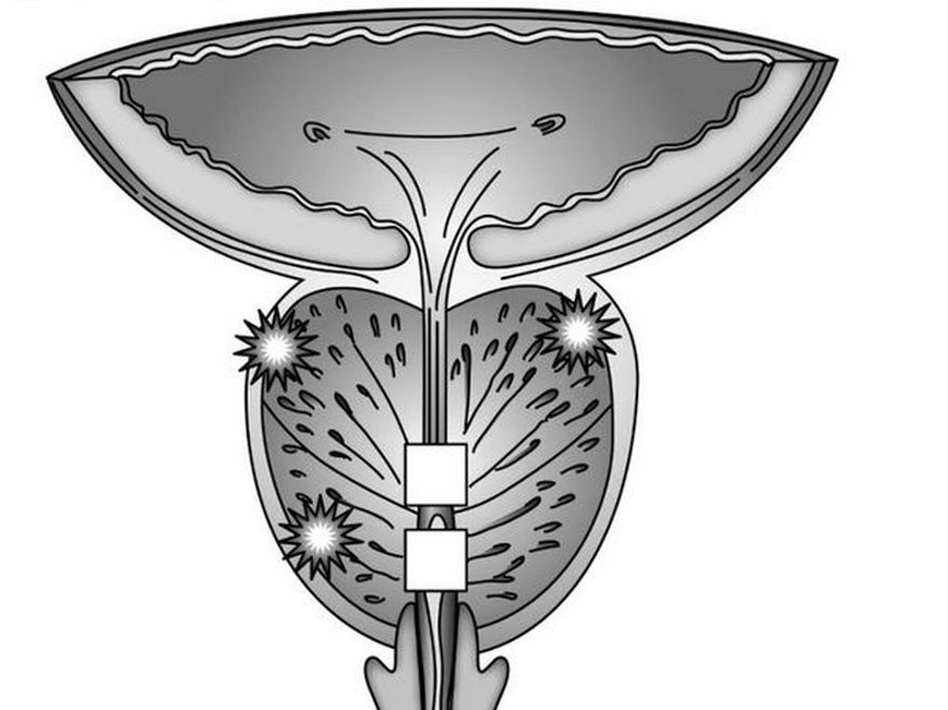
Knowledge of the causes of the development of stones in the prostate gland is necessary for high-quality and adequate etiological therapy, which helps prevent the recurrence of calculous prostatitis.
Symptoms of calculous prostatitis
Calculus prostatitis symptoms develop over a long period of time, and a man may not pay attention to them. The clinical picture of this disease may include symptoms such as dull pain in the lower abdomen and lower back, sacrum, perineum, and pubis.
Pain may begin or intensify after defecation, sexual intercourse, intense physical activity and other provoking factors. Dysuric disorders are observed - a frequent desire to go to the toilet, painful or difficult urination, burning in the urethra and lower abdomen, and sometimes urinary retention occurs due to obstruction in the form of stones.
Patients experience prostatorrhea - involuntary secretion of the prostate gland while resting or during physical exercise, straining during defecation or urination. There may be blood in the urine and semen.
Almost always, against the background of persistent inflammation with the formation of stones, sexual dysfunction develops - weak erection, premature ejaculation, decreased libido.
The main symptoms of calculous prostatitis include:
- erectile dysfunction;
- pain in the groin area, which can be spasmodic and paroxysmal;
- during ejaculation - indicates damage to the prostate tubule channel by the sharp edge of the stone;
- premature and painful ejaculation.

Such symptoms lead to a decrease in sexual desire.
Often men associate this with age factors, mistakenly believing that such sexual dysfunction will not go away. Sometimes they begin to self-medicate using various erectile dysfunction drugs (PDE-5 inhibitors).
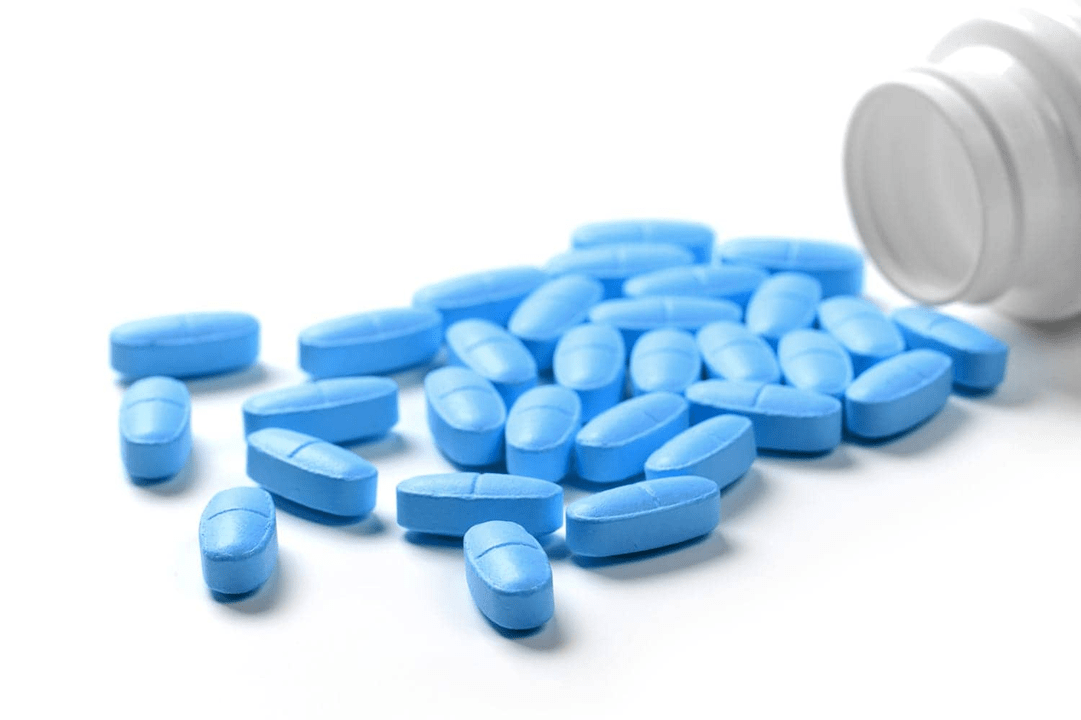
This approach is very dangerous, because it can worsen the course of the pathological process and lead to the development of complications.
Prostatitis is an inflammatory pathological process in the prostate gland of a man. In most cases, it is caused by an infection, which gradually leads to chronic, long-term illness and the development of complications.
Treatment of calculous prostatitis is complex
- antibiotics,
- anti-inflammatory drugs,
- enzymes
- immune drugs
- phytotherapy,
- physiotherapeutic procedures.
Antibacterial agentprescribed as part of etiotropic treatment. Their intake is necessary to block the activity of the causative agent of the infectious inflammatory process. These can be non-specific microbial flora (streptococci, staphylococci, enterococci, Escherichia coli, Proteus), and specific pathogens of urogenital infections - gonococcus, chlamydia, ureaplasma, trichomonas, etc.
The choice of antibiotics may be based on the results of culture studies of prostate secretions and the determination of the sensitivity of microbial pathogens to drugs. Sometimes antibiotics are prescribed empirically based on scientifically proven antimicrobial efficacy of drugs. The selection of antibiotics, the determination of the dosage and the duration of their use can be done exclusively by the attending physician, because their uncontrolled use can lead to serious complications and worsen the course of the underlying disease.
If the tissue of the prostate gland is parasitized by poly-related microbial flora (bacteria, viral microorganisms, protozoa), the etiotropic therapy regimen will consist of a complex of different drugs that act in a specific antimicrobial spectrum.
To stimulate the body's immune defensesand its resistance to infection, immunomodulatory drugs are prescribed - Immunomax, Panavir, Interferon and its derivatives. To increase the antimicrobial effect of etiotropic drugs, enzymatic agents are prescribed along with them - longidase, chemotrypsin. They facilitate the delivery of active antibiotic substances to the affected tissue, have an indirect analgesic effect, and have anti-inflammatory and regenerative effects.
Pain syndrome relieved withuse non-steroidal anti-inflammatory drugs. Along with antibiotic therapy, probiotics are prescribed to prevent the development of intestinal dysbiosis. To protect the liver parenchyma from the toxic effects of antibacterial drugs and improve its functional state, hepaprotectors are prescribed. After the acute inflammatory phenomenon subsides, physiotherapeutic procedures are prescribed - laser treatment, magnetic therapy, mud therapy, galvanization, medical electrophoresis, reflexogenic therapy, hardware treatment, etc.
This improves metabolic processes, microcirculation, lymphatic drainage and prostate tissue trophism, stimulates the restoration of its functional state and helps resolve the inflammatory process. To destroy the stone, a low-frequency laser is used. It breaks up stones and allows small stones to pass out of the tubules. In case of complications in the form of prostate adenoma or abscess (restricted cavity filled with pus), surgical intervention is performed.

It involves the removal of part of the prostate gland (resection). To avoid this, at the first signs of pathology, which is expressed in erectile dysfunction, you need to consult a doctor. Self-medication or ignoring the problem always leads to the development of subsequent complications.


